Cytospora cedri Syd., P. Syd. & E.J. Butler, Annls mycol. 14: 193 (1916)
Index Fungorum number: IF 184521
Associated on branches of Rubus sp. Asexual morph: Pycnidial stromata immersed in the bark, scattered, producing black area on the bark, circular to ovoid, with multiple locules, occasionally slightly erumpent through the surface, ostiolate. Ostiole in the centre of the disc, black, conspicuous. Locules 150–500 μm, numerous, subdivided frequently by invaginations with common walls. Conidiophores hyaline, septate, branched at base, middle, thin-walled, occasionally septate, embedded in a gelatinous layer. Conidiogenous cells 4–7(–8) × 0.8–1.5 μm (x̅ = 7 × 1.4), enteroblastic, phialidic, sub-cylindrical. Conidia (3–)4.5–5 × 0.5–1 μm (x̅ = 5 × 0.8), hyaline, allantoid, aseptate, smooth-walled. Sexual morph: Undetermined.
Material examined – Italy, on branches of Rubus sp., Erio Campesori IT3288, MFLU 17-0835.
GenBank numbers – ITS: MN871816, LSU: MN873004, RPB2: MN871989.
Known distribution (based on molecular data) – Canada, France, Italy, Portugal, Spain, Switzerland, USA (López-Moral et al. 2020, Shang et al. 2020).
Known hosts (based on molecular data) – Abies alba, Acer saccharum, Castanea sativa, Cedrus deodara, Chamaecyparis, Cupressus sempervirens, Lonicera sp., Ostrya carpinifolia, Quercus ilex, Rubus sp., Thuja sp. (López-Moral et al. 2020, Shang et al. 2020).
Notes – Cytospora cedri was introduced by Sydow et al. (1916) from Cedrus libani in India. Our strain was identified as C. cedri based on morphological characteristics and phylogenetic analyses (Fig. 36). The conidiophore and conidial characters of the type species differ slightly with our isolate by having allantoid conidia of 4–6 × 0.5–1.2 µm and 10–12 × 1–1.3 µm conidiophores (Sydow et al. 1916).
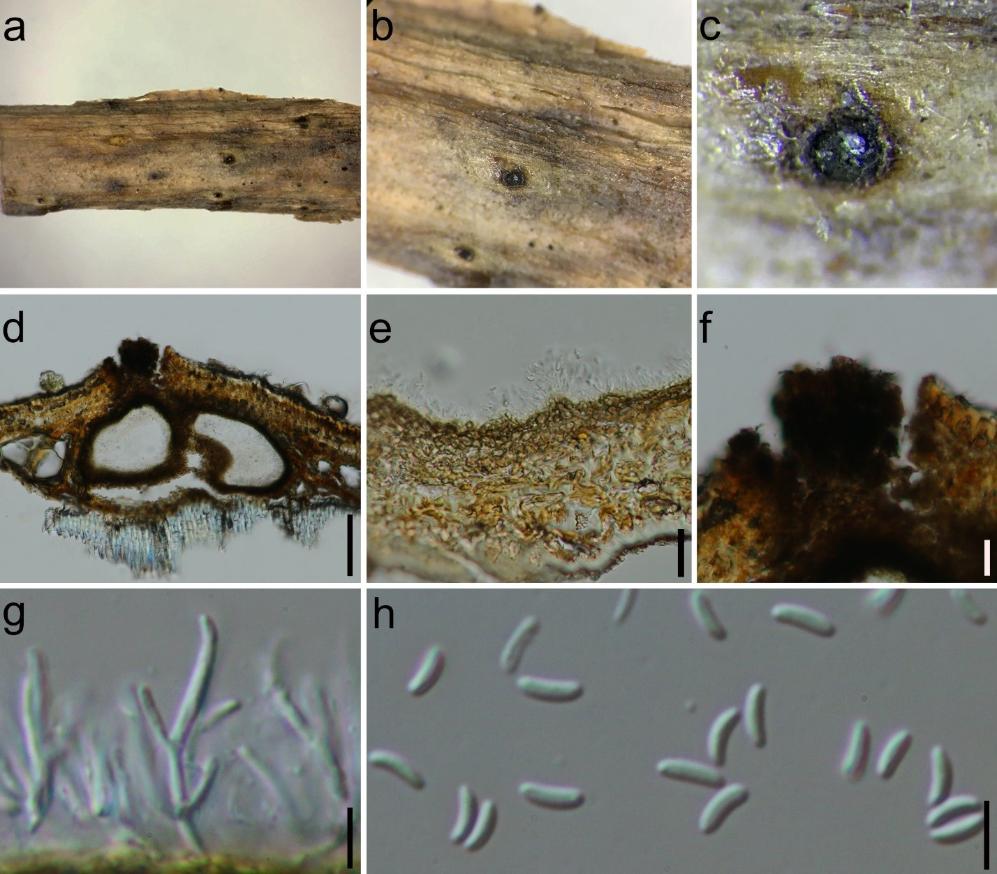
Figure 1. Cytospora cedri (MFLU 17-0835, new host record). a Stromatal habit in wood. b Fruiting bodies on host surface. c Surface of fruiting bodies showing the black ostioles. d Cross section of the stroma showing conidiomata. e Peridium. f Ostiolar neck. g, h Conidiogenous cell containing conidia. i Conidia. Scale bars: d = 100 µm, e, f = 20 µm, g–i = 5 µm.