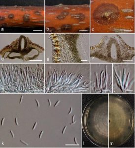
Cytospora ampulliformis Norphanphoun, Bulgakov, T.C. Wen & K.D. Hyde, sp. nov., Index Fungorum number: IF552601
Etymology: The specific epithet ‘ampulliformis’ refers to the flask-shaped conidiogenous cells.
Associated with twigs and branches of Sorbus intermedia (Ehrh.) Pers. Sexual morph: Undetermined. Asexual morph: Conidiomata 680–1200 × 350–480 μm diameter, semi-immersed in host tissue, solitary, erumpent, scattered, discoid, circular to ovoid, with 3–4 locules, and ostiolar neck. Ostioles 200–300 μm long, at the same level as the disc surface. Peridium comprising few to several layers of cells of textura angularis, with inner most layer thin, hyaline to pale brown, outer layer brown to dark brown. Conidiophores unbranched, reduced to conidiogenous cells. Conidiogenous cells blastic, enteroblastic, flask-shaped phialidic, formed from the inner most layer of pycnidial wall, hyaline, smooth-walled. Conidia (5–)5.6–9 × 1.3–1.6(–1.7) μm (x̅ = 7.5 × 1.6 μm, n = 30), unicellular, subcylindrical, hyaline, smooth-walled.
Culture characteristics: Colonies on MEA, reaching 7.5 cm diameter after 7 days at 25 °C, producing dense mycelium, circular, margin rough, white, lacking aerial mycelium.
Material examined: RUSSIA, Rostov Region, Rostov-on-Don City, Botanical Garden of Southern Federal University, Systematic Arboretum, parkland, dead and dying branches (necrotrophic) of Sorbus intermedia (Rosaceae), 30 May 2015, T. Bulgakov, T-483 (MFLU 15-2187, holotype, KUN, isotype), ex-type living culture, MFLUCC 16-0583, KUMCC; RUSSIA, Rostov Region, Krasnosulinsky District, Donskoye Forest, artificial forest, dying twigs and branches (necrotrophic) on Acer platanoides L. (Sapindaceae), 27 October 2015, T. Bulgakov, T-1094 (MFLU 15-3756, KUN), living culture, MFLUCC 16-0629, KUMCC.
Notes – Cytospora species associated with Sorbus sp. were reported in previous studies such as C. leucostoma and Valsa massariana (Adams et al. 2002, 2005). In this study, C. ampulliformis, C. sorbi and C. sorbicola are also reported from Sorbus sp. (Table 3). Morphologically, C. ampulliformis is similar to C. sorbi in its conidiomata having 3–4 locules and in the length of ostiolar neck (C. ampulliformis: 200–300 μm versus 250–300 μm: C. sorbi). However, C. ampulliformis differs from C. sorbi in having larger conidia (C. ampulliformis: 7.5 × 1.6 μm, versus 6.5 × 1.5 μm: C. sorbi). Based on phylogenetic analyses, both species form separate lineages within the genus Cytospora .
We obtained two isolates of Cytospora ampulliformis (MFLUCC 16-0583, MFLUCC 16-0629) and these formed a close relationship with C. cotini (MFLUCC 14-1050) isolated from Cotinus coggygria, C. tanaitica (MFLUCC 14-1057) isolated from Betula pubescens, C. rosarum (218) isolated from Rosa canina and C. ulmi (MFLUCC 15-0863) isolated from Ulmus minor. Cytospora ampulliformis (7.5 × 1.6 μm) differs from C. cotini in forming black-discoid conidiomata on the host and lobate circular colonies on MEA (Hyde et al. 2016), while it differs from C. cotini (5.9 × 1.2 μm), C. tanatica (3.4 × 0.7 μm), C. rosarum (5–6 × 1.5 μm) and C. ulmi (5.4 × 1.4 μm) by its larger conidia.

