Cryptocalicium blascoi Etayo, Olariaga and M. Prieto, sp. nov.
MycoBank number: MB 837519; Index Fungorum number: IF 837519; Facesoffungi number: FoF;
Holotypus: Spain, Madrid, Hoyo de Manzanares, Parque de la Cabilda, 40.626832 -3.897757, on the inner side of loose barkstrips of Juniperus oxycedrus, 15 May 2020, leg. I. Olariaga, ARAN-Fungi 14723. Ex-type culture: CECT 21190.
Etymology: named after Javier Blasco Zumeta, outstanding Aragonese naturalist, who showed to us the first locality where this species was found and who provided us with further material of it.
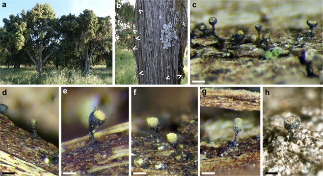
Ascomata saprobic on the inner side of loose bark strips (Fig. 1A–B) still attached to the tree, solitary or gregarious, 0.15–0.36 mm high (mazaedium excluded) (Fig. 2 C–H), Fig. 3A). Capitulum urniform to hemispherical, 0.05–0.10 × 0.10–0.15 mm, dark violaceous brown when wet, black and shiny when dehydrated, monocephalic, with an ochre disc. Mazaedium well developed, light ochre (4A6) to greyish green (1D2), spores that keep together. Stalk cylindrical, sometimes with a broader base, straight, seldom bent, smooth, black, shiny, 0.10–0.26 × 0.02–0.04 mm.
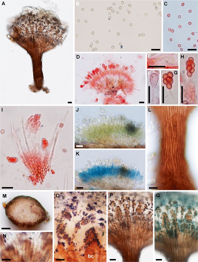
Ascospores (Fig. 3B) globose to subglobose, rarely ellip-soid, hyaline inside asci, pale brown when mature, with homogeneous content, with two minute refractive bodies in Congo Red (Fig. 3C), smooth when viewed in a light micro- scope, (3)3.3–4(4.7) × 2.8–3.5 (4.0) μm (Lm=3.7–3.9, Wm=3.2–3.3, Q=1.08–1.25(1.36), Qm=1.15–1.19; n=25),
slightly thick-walled (0.2–0.4 μm). Asci clavate, bitunicate, initially thick-walled (wall 1 μm), then thin-walled, evanescent, 8-spored, 20–27 μm long; pedicel 1 μm diam.; sporiferous part 10 – 16 × 5 – 7 μm ( Fig. 3D– H). Hamathecium composed of regularly scattered sterile elements, protruding 10–30 μm over mature asci, cylindrical, obtuse, with 2–4 septa, hyaline to very pale brown, smooth or slightly rough, 32–40 × 1.5–2 μm (Fig. 3I). Subhymenium poorly differentiated from the medullary excipulum, com- posed of a textura angularis-globulosa with more or less isodiametrical hyphae, pale brown in water, green in IKI (Fig. 3J) and amyloid in KOH+IKI (Fig. 3K). Stalk corticate (Fig. 3 L–M). Outermost layer of the stipe composed of 1–2 rows of parallel-arranged hyphae, cylindrical, aseptate, slight- ly thick-walled, dark reddish brown, tightly adhered, un- changed in KOH, 2.7–3.5 μm broad. Stalk medulla composed of parallel-arranged hyphae, cylindrical, septate, hyaline, thick-walled (0.2–0.4), 1.2–1.6 μm broad. Ectal excipulum poorly differentiated from the medullary excipulum, com- posed of periclinal-arranged hyphae, of textura prismatica in surface view (Fig. 3N), with cells progressively shorter towards the margin of the capitulum, pale brown, thick- walled (0.5–1 μm), 3.5–5.5 μm broad, covered with crusts of dark reddish brown pigment (Fig. 3O), unchanged in KOH. Medullary excipulum 15–20 μm thick, composed of a textura angularis-globulosa with more or less isodiametrical hyphae, hyaline to very pale brown, thin-walled, 2–4 μm broad, non-gelatinized. Pigment granules present on the ectal excipulum (Fig. 3O–P), surface of the stipe and the subhymenium, dark violet, opaque, partly dissolving and turning turquoise green in KOH (Fig. 3Q).
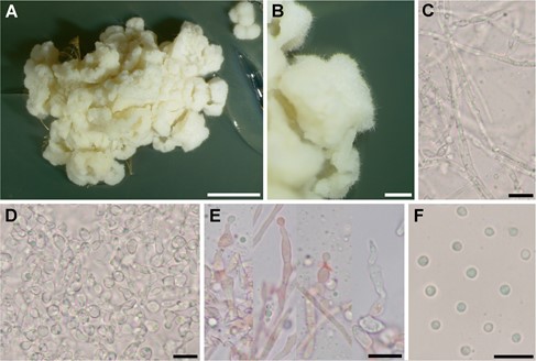
Colonies on MEA 9–17 mm diam and 4–6 mm high after 90 days at 5 °C, superficial, effuse, convex to obtusely conical, initially even, later wrinkled-cerebriform, cauliflower- like, cream white, tomentose (Fig. 4A). Reverse pale brownish ochre. Margin lobed, distinct (Fig. 4B). Vegetative hyphae sometimes arranged in parallel, cylindrical, septate, some- times constricted at septa, with sparse intercalary connections, hyaline, smooth, 1.5–2 μm broad (Fig. 4C). Asexual morph hyphomycete-like, observed only in culture. Conidiophores 30–48 μm long, covering the surface of cultures, basally branched, formed by 2–6(8) catenulate segments (Fig. 4D). Segments irregular in shape, usually cylindrical to subglobose, often triangular or angled, constricted at septa and connected to each other by 1–3, thin, 1 μm broad appendices, easily disarticulating in microscopic preparations, 5–9× 3–6.5 μm (Fig. 4E). Conidiogenous cells cylindrical to ovoid, 5–12 × 1.5–4 μm, bearing one simple apical conidiogenous locus, 1 μm broad, to which immature conidia are attached. Conidia single, first pyriform then subglobose, slightly truncate at the basal end, thin-walled, smooth, hyaline, with small oily droplets, 2.3–3 μm (Lm = 2.6) (Fig. 4F).
Additional specimens examined: SPAIN. Ávila: El Barraco, south shore of Embalse del Burguillo, Peña del Roble, 40.409325 -4.567302, on bark strips of Juniperus oxycedrus, 3 October 2020, M. Prieto & I. Olariaga, ARAN- Fungi 14748. Burgos: Briongos de Cervera, espacio natural La Yecla y Sabinares de Arlanza, stand with large J. thurifera trees, 300 m ahead the town, 41.913889 -3.497222, 1095 m, on bark strips of Juniperus thurifera, 04 August 2019, J. Etayo 31925 (MAF). Santo Domingo de Silos, track from Carazo, espacio natural La Yecla y Sabinares de Arlanza, km 48, 1130 m, on bark strips of J. thurifera, 41.958333 – 3.400000, 04 August 2019, J. Etayo 31836 (hb. Etayo). Madrid: Hoyo de Manzanares, Camino del Cementerio, 40.623941 -3.898383, 20 m from the crossing with M-618 road, on bark strips of Cupressus sempervirens, 4 June 2020, I. Olariaga, ARAN-Fungi 14725. Moralzarzal, La Navata, near the Arroyo de Peregrinos stream, 40.617972 -3.951778, on loose bark strips of Juniperus oxycedrus, 7 June 2020, M. Prieto and I. Olariaga, ARAN-Fungi 14724. Pedrezuela, El Tiradero, 40.740483 -3.637523, on bark of a large J. oxycedrus tree, 27 March 2021, M. Prieto & I. Olariaga, ARAN-Fungi 15628. Soto del Real, close to Arroyo del Recuenco, 40.754794 -3.834830, on the underside of bark strips of Juniperus oxycedrus, on dried resin drops, 13 June 2020, M. Prieto and I. Olariaga, ARAN-Fungi 14726. Soria: Borobia, El Frontón, 41.646448 -1.913891, on bark strips of Juniperus thurifera, 17 October 2020, I. Olariaga, ARAN-Fungi 15627. Toledo: Almorox, near arroyo de Labros, 40.300343 -4.243062, on bark strips of Juniperus oxycedrus, 10 October 2020, M. Prieto and I. Olariaga, ARAN-Fungi 14747. Zaragoza: Pina de Ebro, Retuerta de Pina, on underside of bark strips of Juniperus thurifera, 9 October 2017, J. Blasco, Etayo 30875, 31798 (hb. Etayo).

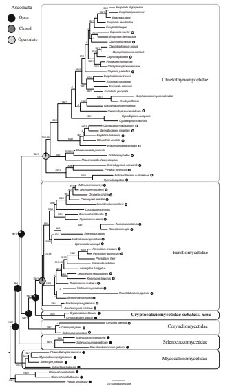
Fig. 1 Best maximum likelihood tree obtained from RAxML based on a 7-locus data set (5.8S nuITS, nuLSU, nuSSU, mtSSU, MCM7, RPB1 and RPB2). Nodes show bootstrap support (ML-BS) from maximum likelihood, and posterior probabilities (PP) obtained in the Bayesian analysis, ordered as ML-BS/PP. ML-BS below 60% and PP below 0.85 are not shown. Results from the ancestral state reconstruction with BayesTraits are shown in the studied nodes. Ascoma types are also indicated except for those species devoid of sporocarps or with unknown sexual morphs

Fig. 2 Cryptocalicium blascoi (holotype). A Type locality. B Juniperus oxycedrus tree with loose bark strips. White arrows indicate suitable places for C. blascoi. C Gregarious ascomata. D Young ascoma with unexposed hymenium. E Older ascoma with exposed hymenium. F–G Ascomata with mazaedia. H Old ascoma without mazaedium (Etayo 31798). Scale bars = 100 μm. Photographs I. Olariaga

Fig. 3 Microscopic features of Cryptocalicium blascoi (holotype unless otherwise indicated). A Ascoma (Etayo 31798). B Ascospores in water. C Ascospores in CR. D Portion of hymenium showing asci and protruding sterile elements in SDS-CR. E Young thick-walled ascus in CR. F Older thin-walled ascus in CR. G Mature ascus without wall in CR. H Mature ascus attached to the subhymenium in CR. I Protruding sterile elements (CR). J Hymenium in IKI showing a green reaction. K The same hymenium portion in KOH+IKI, showing an amyloid reaction. L Stalk surface in water. M Stalk section in water. N Cells of the ectal excipulum in surface view in water. O Close-up of the ectal excipulum covered with crusts of dark reddish brown pigment and dark violet pigment granules in water. P Dark violet pigment granules in water (Etayo 31798). Q Partly dissolved and turquoise green pigment granules in KOH (Etayo 31798). Scale bars = 10 μm. Abbreviations: CR, SDS Congo Red; IKI, Lugol solution; KOH, Potassium hydroxide; bc, brown crust. Photographs I. Olariaga

Fig. 4 Cultural characters of Cryptocalicium blascoi (ex- holotype culture CECT 21190). A Colony in MEA. B Colony margin and surface. C Vegetative hyphae in water. D Colony surface showing catenulate and branched conidiophores and some conidiogenous cells and conidia in water. E Conidiogenous cells with conidia in formation in CR. Scale bars = 5 mm (A), 1 mm (B) and 10 μm (C–F). Abbreviations: CR, SDS Congo Red
