Coniella Höhn., Ber. Dt. Bot. Ges. 36(7): 316 (1918)
MycoBank number: MB 7753; Index Fungorum number: IF 7753; Facesoffungi number: FoF 04309; 57 morphological species (Species Fungorum 2020), 38 species with sequence data.
Type species – Coniella fragariae (Oudem.) B. Sutton
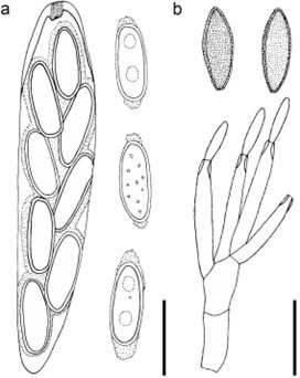
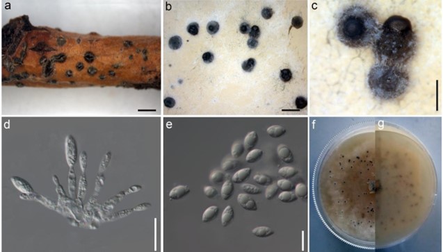
Notes – The cosmopolitan genus, Coniella was established by Höhnel (1918a) and is typified by Coniella pulchella (= Coniella fragariae). Species of this genus are plant pathogens (van Niekerk et al. 2004, Mirabolfathy et al. 2012, Chethana et al. 2017). In addition, they have saprobic, endophytic, and parasitic lifestyles, as well as being secondary invaders of plant tissues infected by other organisms or injured by other causes (Samuels et al. 1993, Ferreira et al. 1997, Alvarez et al. 2016). Several Coniella species are host-specific, contradictory to their wide host range (Alvarez et al. 2016, Chethana et al. 2017). This genus has been subjected to many revisions. Initially, Coniella and Pilidiella were given distinct identities due to their conidial pigmentation (von Arx 1973, 1981b). Conidial pigmentation was rejected as a distinguishing character and Pilidiella was synonymized under Coniella (Sutton 1980, Nag Raj 1993). With molecular data used in species delimitation, Coniella and Pilidiella were considered as distinct genera (Castlebury et al. 2002, van Niekerk et al. 2004, Wijayawardene et al. 2016b). In an attempt to resolve many species complexes residing in the genus, Alvarez et al. (2016) accepted Coniella as the only genus in Schizoparmaceae and synonymized Pilidiella and Schizoparme. The most recent phylogenetic status of this genus is by Chethana et al. (2017). Coniella destruens and Coniella vitis are illustrated in this study (Figs. 225, 226).

Figure 225 – Coniella destruens (redrawn from van Niekerk et al. 2014). a Ascus and ascospores of Coniella destruens. b Conidiophores and conidia of C. destruens. Scale bars: a, b = 10 µm.

Figure 226 – Coniella vitis (Material examined – RUSSIA, Rostov Region, Shakhty City, on dead and dying branch of Salix alba L. (Salicaceae), 5 March 2016, T.S. Bulgakov, T1278 (MFLU 16– 1572). a Host tissue. b Submerged pycnidia on PDA. c Close view of the pycnidia and the spore mass. d Conidiogenous cells. e Hyaline to brown conidia. f, g Upper view (f) and the reverse view (g) of the colony on the PDA. Scale bars: a, b = 1 mm, c = 500 µm, d = 5 µm, e = 10 µm.
Species
Coniella fragariae
