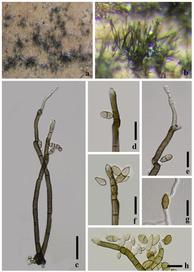
Cladosporium pseudocladosporioides Bensch, Crous & U. Braun, Studies in Mycology 67: 71 (2010). Fig. 7
MycoBank number: MB 517087; Index Fungorum number: IF 517087; Facesoffungi number: FoF 06968;
Saprobic on Nelumbo sp. Colonies on natural substrate effuse, black, velvety. Sexual morph: Undetermined. Asexual morph: Hyphomycetous. Mycelium partly immersed, partly superficial, composed of branched, septate hyphae. Conidiophores up to 250 μm long, 4.0–7.5 μm wide, macronematous, mononematous, erect, straight to slightly flexuous, cylindrical, brown, paler towards apex, hyaline at apex, unbranched, septate, not constricted in the septum, smooth-walled, thick-walled. Conidiogenous cells 16–38 × 4–6 μm, polyblastic, integrated, terminal, subhyaline to pale brown, cylindrical, subdenticulate. Conidia 7–20 × 4–6.5 μm (x̅ = 12 × 5 μm, n = 30), catenate, small terminal conidia globose, subglobose to obovoid, subhyaline to pale brown, aseptate, broadly rounded at the apex; intercalary ramoconidia conidia subglobose, broadly ellipsoid-ovoid, pale brown to median brown, 0–3 septate, with distal hila.
Material examined – China, Guizhou, Xingyi, Anlong, on leaves of Nelumbo sp. (Nelumbonaceae), 27 October 2017, Yao Feng, AL-7 (GZAAS 20-0006), living culture GZCC 20-0010.
Culture characteristics – Conidia germinating on water agar media within 24 h. Germ tubes produced from one or both ends. Colonies on PDA circular, edge entire, mycelia dense, greyish brown from above, dark brown from below.
Notes – Bensch et al. (2010) introduced C. pseudocladosporioides. This species has been reported worldwide on diverse hosts, as well as isolated from air and soil (Bensch et al. 2010, 2012, 2018). Cladosporium pseudocladosporioides belongs to C. cladosporioides complex (Bensch et al. 2015). In the phylogenetic analyses (Fig. 8), our strain formed a strongly supported clade with eight C. pseudocladosporioides strains.

Figure 7 – Cladosporium pseudocladosporioides (GZAAS 20-0006). a, b Colonies on natural substrate c, h Conidiophores and conidia d–f Conidiogenous cells g Germinated conidium Scale bars: c = 30 μm, d–h = 15 μm.
