Chaetothyrina mangiferae Singtripop, Hongsanan & K.D. Hyde, sp. nov.
Etymology: The epithet mangiferae’ refers to host, Mangifera indica
Holotype: MFLU 14-0191.
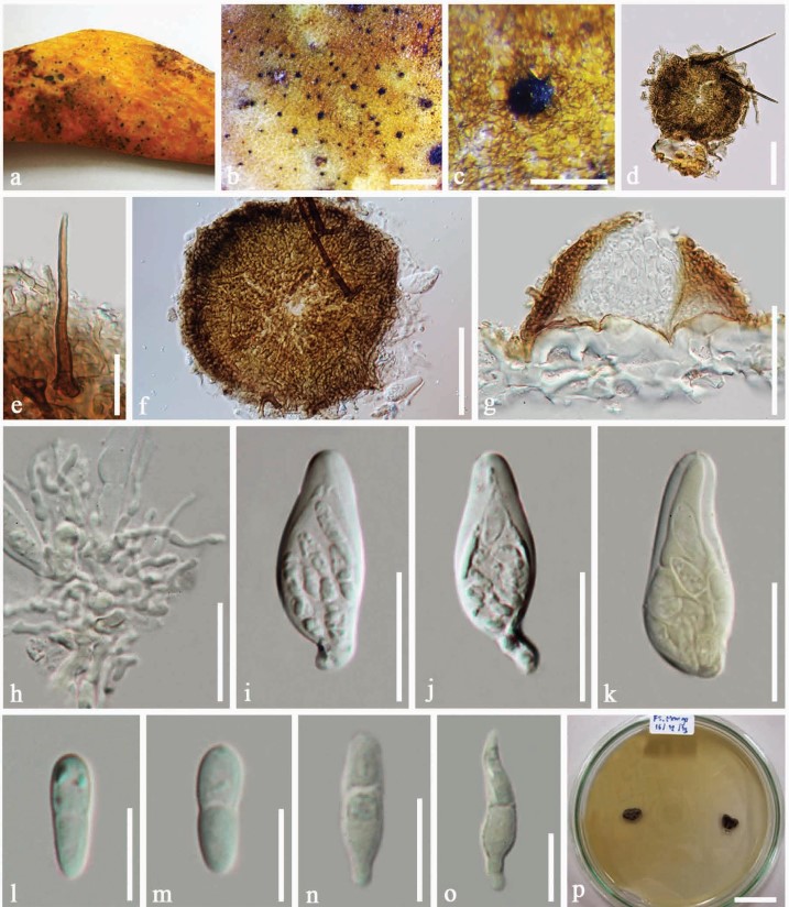
Epiphytes associated with the sooty blotch and flyspeck complex disease of fruit of Mangifera indica, appearing as very small, black, shiny dots, round to ovoid, with long setae. Sexual morph: Thyriothecium 90–130 × 80–120 µm (x̄=110 × 104 µm, n=5), superficial or partly immersed in host tissues, solitary, circular, brown to dark brown, rounded and darkened at the margin, easily removed, base poorly developed, with setae, ostiole central, lacking superficial mycelium. Setae 50-120 µm long, arising from the surface of thyriothecium, straight, rounded at the apex, unbranched, septate, darkened at the septa, brown to dark brown, smooth-walled. Upper wall comprising 2-3 layers of brown to dark brown cells of textura epidermoidea. Hamathecium of 3-5 µm wide, septate, hyaline pseudoparaphyses. Asci 26–36 × 11–12 µm (x̄=32 × 12 µm, n=5), 8-spored, bitunicate, broadly oblong to pyriform, ends apically rounded, with an ocular chamber. Ascospores 11–23 × 3–6 µm (x̄=16 × 5 µm, n=10), 2–3-seriate, hyaline, broadly ellipsoidal to fusoid, 1-septate, slightly constricted at the septa, upper cell mostly larger than lower cell, rounded ends, smooth-walled. Asexual morph: Undetermined.
Culture characters: Ascospores germinating on MEA at 25–28 °C after 24–36 hours in the dark, germ tubes appearing from each end of the ascospores. Colonies reaching 0.8–1 cm diam. after 15 days on MEA at 25–28 °C raised, comprising raised dark grey mycelium, white to greyish at the margin.
Material examined: THAILAND. Chiang Rai Province: Tar Sud District, on living fruit of Mangifera indica (Anacardiaceae), 7 December 2013, Chonticha Singtripop FS01 (MFLU 14-0191, holotype, HKAS 90977, isotype); Tar Sud District, on living fruit of M. indica, 18 January 2015, Chonticha Singtripop (MFLU16-0886), ex-type living culture, MFLUCC 14-0201, MUCL55902.
Notes: Chaetothyrina mangiferae is most similar to C. applanata (Ellis & G. Martin) M.E. Barr, in having superficial scutate thyriothecia with dark brown, septate setae, an upper wall comprising cells of textura epidermoidea, bitunicate, oblong to pyriform asci and 1-septate, hyaline ascospores (Barr 1993), However, it differs in having smaller thyriothecia, larger, ellipsoidal to fusoid ascospores and a different host.
Fig. 4. Chaetothyrina mangiferae (holotype). a, b Disease symptoms on fruit. c Appearance of thyriothecium on host. d Thyriothecium when viewed in squash mount. e Seta arising from the surface of thyriothecium. f Upper wall of thyriothecium when viewed in squash mount. g Section of thyriothecium. h Pseudoparaphyses. i, j Asci. k Ascus in Melzer’s reagent. l, m Ascospores. n, o Ascospores germinating in Melzer’s reagent. p Colonies on MEA plate viewed from above. Scale bars: b=1,000 µm, c=200 µm, d=100 µm, f, g=50 µm, e, h, k=20 µm, l-o=10 µm, p=2 cm.