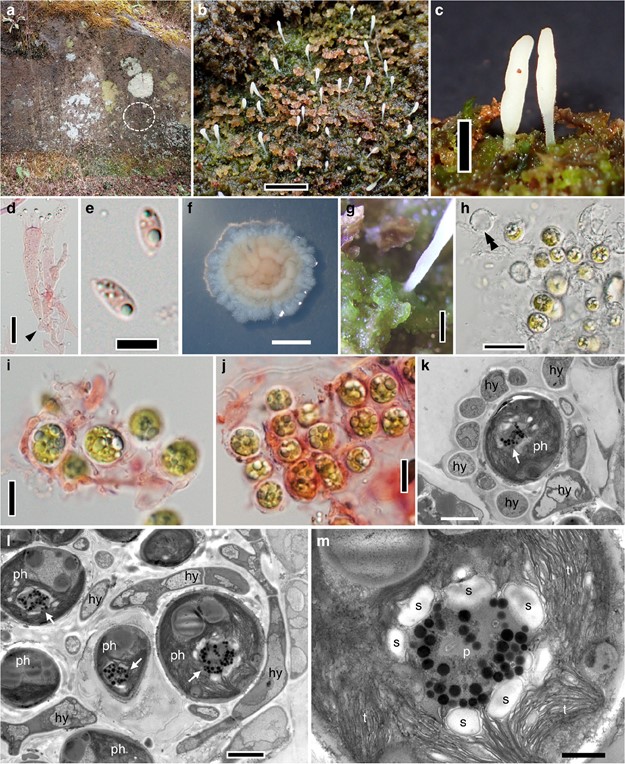
Bryoclavula phycophila H. Masumoto & Y. Degawa, sp. nov. (Fig. 2)
MycoBank number: MB 833864; Index Fungorum number: IF 833864; Facesoffungi number: FoF;
Etymology: phycophila = phyco (Greek) + philus (Latin), referring to “algal” + “loving”
Holotype: JAPAN, Kumamoto, Kikuchi, Kikuchi Valley (33.001070 N; 130.948191 E), on unidentified senescent bryophytes growing on a moist rock surface, elevation 564 m, 18 Dec 2018, Hiroshi Masumoto coll. no. 293 (TNS- F-79667, isotype: OSA-MY-9328, ex-type culture: NBRC 114394).
Gene sequences of the holotype: LC508117 (ITS1-5.8S- ITS2), LC508118 (nrLSU).
Gene sequences of the ex-type culture: LC544111 (ITS1- 5.8S-ITS2), LC544112 (nrLSU).
Description: Thallus formed on unidentified senescent bryophytes growing on a moist rock surface, gelatinous, bright green, amorphous, undifferentiated, composed of spherical photobiont cells loosely surrounded by mycobiont hyphae, not developed to form a globular thallus like Multiclavula species. Photobiont green algae, cells subglobose, 8.2–13.8 μm in diam, containing a pyrenoid and several oil droplets, each algal cell loosely surrounded by mycobiont hyphae of 1.9–3.0 μm thick. Basidiomata dispersed on unidentified senescent bryophytes growing on a moist rock surface, fleshy, simple, rarely branched, clavate or fusiform, 1.6–3.8 mm high, 0.2–0.6 mm wide, whitish to pale cream, apex broadly to narrowly rounded, with a basal stipe, 0.9–2.8 mm high, 140–300 μm wide, usually less than half the length of basidiomata, subtranslucent. Trama hyphae 2–6 μm in diam, parallel aligned, hyaline, clamped, then- to slightly thick-walled ( wall up to 0 . 6 μm thick), densely agglutinated. Hymenium 55–90 μm thick. Basidia 25–50 × 5–8 μm, cla- vate to suburniform, thin-walled, hyaline, with a basal clamp connection, with (2)4–6(7) sterigmata, 2.2–5.0 μm long. Basidiospores (4.4)5.6 – 6. 6(7.5) × ( 2 . 4)2.7– 3.2(3.6) μm [av. ± std. 6.09 ± 0.54 × 2.94 ± 0.25 μm, n = 150], Q = (1.5)1.9–2.3(2.7) [av. ± std. 2.09 ± 0.23, n = 150], narrowly ellipsoid to elongate, hyaline, smooth, thin-walled, inamyloid, usually guttulate. Colonies on PDA slow growing, reaching 1.1–1.3 cm in diam after 2 months at 20 °C in the dark, velvety, cream to pale brown in center, whitish in outer part, with undulate margin.
Habitat and ecology: The new taxon occurs on unidentified senescent bryophytes on a moist large rock outcrop. Although this fungus forms basidiomata on bryophytes, no direct relationship between the mycelia and the bryophytes was observed. The mycelia are consistently associated with green algae present on the surface of the bryophytes, indicating the lichenization of this species. The basidiomata were collected in late autumn to early winter (November and December). In addition to this species, Dibaeis absoluta (Tuck.) Kalb & Gierl occurred on the same rock outcrop.
Distribution: This species is so far known only from the type locality (Kumamoto) located in the temperate zone in Japan.
Additional specimen examined: JAPAN, Kumamoto, Kikuchi, Kikuchi Valley (33.001070 N; 130.948191 E), on bryophytes growing on a moist rock, elevation 564 m, 20 Nov 2018, Yousuke Degawa (paratype: TNS-F-79666).
Gene sequences of the paratype: LC544109 (ITS1–5.8S- ITS2), LC544110 (nrLSU).
Notes: As a result of the observation on the lichenized thallus of Bryoclavula phycophila by LM and TEM, the hyphae loosely surrounded each algal cell (Fig. 2h, i) but did not enclose multiple algal cells to form the globular thallus-like Multiclavula. No haustoria were observed. Though it is un- clear that the photobiont has a pyrenoid by LM, TEM observation clearly revealed that the photobiont cell has a pyrenoid associated with starch grains (Fig. 2k–m).

Fig. 2 Bryoclavula phycophila (TNS-F-79667, holotype). a Habitat, a large rock outcrop. The dotted circle represents the area where B. phycophila occurred. b Basidiomata on senescent bryophytes growing on a moist rock surface. c Details of basidiomata with a subtranslucent stipe. d Basidium with a basal clamp connection (arrowhead). e Basidiospores. f Culture (NBRC 114394) grown on PDA for two months at 20 °C. g Amorphous thallus. h–j Details of thallus. A Double arrowhead indicates a hyphal ring that once surrounded the photobiont cell. k, l TEM images of the thallus. Photobiont cells contains a pyrenoid (arrows). m TEM image of a pyrenoid of the photobiont cell. d, e, i, and j are mounted in 3% KOH and stained with 1% Congo red. hy = hyphae, p = pyrenoid structure, ph = photobiont cells, s = starch grains, t = thylakoid membrane. Scale bars: b, f = 5 mm; c = 1 mm; g = 500 μm; h = 20 μm; d, i, j = 10 μm; e =5 μm; k, l =2 μm; m = 500 nm
