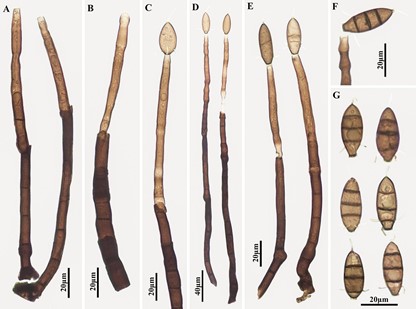
Blastophragmia plurisetulosa Jian Ma, L.G. Ma, X.G. Zhang & R.F. Castañeda, sp. nov. Fig. 1
MycoBank number: MB 557507; Index Fungorum number: IF 557507; Facesoffungi number: FoF;
Differs from Endophragmiella spp. by its setulate conidia; and from Stratiphoromyces spp. by its conidiophores either determinate or with several enteroblastic percurrent extensions, and its uncurved, fusiform to ellipsoidal, 3-euseptate conidia with a single apical setula and 2–4 basal setulae.
Type: China, Hainan
Province: Diaoluoshan Mountain, 18°4’N 109°53’E, on dead branches of an unidentified broadleaf tree, 15 April 2015, J. Ma (holotype, HJAUP M0349).
Etymology: refers to the multiple setulae, which arise from the conidial base and apex.
Colonles on the natural substrate effuse, brown to dark brown, hairy. Mycelium partly superficial, partly immersed in the substratum, composed of branched, septate, pale brown to brown, smooth-walled hyphae. Conidiophores macronematous, mononematous, unbranched, erect, straight or flexuous, cylindrical, smooth, 3–16-septate, brown to dark brown, paler towards the apex, determinate or with 1–3 enteroblastic percurrent extensions, ≤340 µm long, 6.5–13.5 µm wide. Conidiogenous cells monoblastic, integrated, terminal, cylindrical, brown to pale brown, smooth, 20–105 × 5.5–6.5 µm. Conidial secession rhexolytic. Conidia solitary, dry, acrogenous, fusiform to ellipsoidal, smooth, brown, 3-euseptate when mature, 25–32.5 × 10.5–12.5 µm; with a single apical setula and 2–4 basal setulae, thin, 5–10 µm long, straight to curved, hyaline; bearing a distinct 3.5–5 µm wide basal frill.

Fig. 1. Blastophragmia plurisetulosa (holotype, HJAUP M0349). A, B. Conidiophores showing conidiogenous cells and enteroblastic percurrent extensions, and (in B) conidiophore apex showing an initial conidium. C–E. Conidiophores and conidiogenous cells with immature conidia; F. Conidium seceding rhexolytically from conidiogenous cell; G. Conidia.
