Aurifilum marmelostoma Begoude et al., in Begoude et al., Antonie van Leeuwenhoek 98(3): 273 (2010).
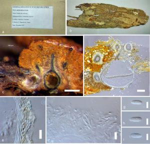
Pathogenic on woody bark. Sexual morph: Ascostromata 300–830 × 760–1050 μm, on bark, gregarious or single, often confluent in cracks, medium to large, ascostromata extending usually beneath or erumpent through bark, semi-immersed, pulvinate to pyriform, orange, upper region eustromatic, lower region pseudostromatic, pseudoparenchymatous to prosenchymatous tissue. Ascomata 190–310 × 170–275 μm, perithecial, valsoid, up to nine per stroma, embedded in stroma at irregular levels with bases touching host tissue, fuscous black, globose to subglobose. Perithecial necks 30–100 μm wide, periphysate, black, emerging at stromatal surface as black ostioles, surrounded with orange stromatal tissue to form papillae, textura porrecta. Peridium 7–12 μm (x̅ = 10 μm, n = 20), comprising inner, hyaline, compressed, cells of textura angularis and outer, thick-walled, brown cells of textura angularis. Asci 50–55 × 7.5–9 μm, 8-spored, unitunicate, fusoid to ellipsoidal, floating freely in the perithecial cavity, unitunicate with non-amyloid, refractive apical rings, non-stipitate. Ascospores 10–12 × 3–4 μm, hyaline, fusoid to ellipsoidal, one median septum with tapered apex. Asexual morph: Conidiomata up to 660 μm high, 600 μm in diam., part of ascomata as conidial locules or as solitary structures, orange, necks absent, tissue around ostiolar openings darkened, broadly convex, semi-immersed, uni- to multilocular, even to convoluted lining. Locule 80–300 μm diam., tissue mostly prosenchymatous with pseudoparenchyma towards the margin depending on the developmental stage of the structure. Conidiophores 15–40 μm long, cylindrical, aseptate, hyaline. Conidiogenous cells 2.5–3.5 μm wide, phialidic, sometimes with inflated bases, collarettes inconspicuous with attenuated apexes. Paraphyses 30–65 × 2.5–3.5 μm long, sterile, cylindrical. Conidia 3.5–4.5 × 1–1.5 μm, minute, hyaline, cylindricalto allantoid, aseptate, exuded through opening at stromatal surface as orange droplets or tendrils (description based on Begoude et al. 2010).
Material examined: CAMEROON, Mbamalyo, bark of Terminalia ivorensis A. Chev. (Combretaceae), December 2007, Begoude and J. Roux, PREM 60256, holotype.
Notes: The monotypic genus Aurifilum was introduced and typified by Aurifilum marmelostoma. This genus is distinguished from other genera of Cryphonectriaceae in having a distinct asexual morph broadly comprising convex
conidiomata, presence of darkened ostiolar openings at the apex of the conidiomata, paraphyses or sterile cells longer than 90 μm, one septate, fusoid to ellipsoid ascospores and minute, cylindrical conidia. This is a serious canker causing pathogen on Terminalia species in West Africa which is a popular source of timber, medicine, spiritual and social benefits to rural populations.
Fig. Aurifilum marmelostoma (PREM 60256). a Packet of herbarium. b Herbarium specimen. c, d Vertical cross section of ascomata. e Peridium. f Asci. g–i Ascospores. Scale bars: c = 200 μm, d = 100 μm, e = 10 μm, f = 20 μm, g–i = 5 μm.

