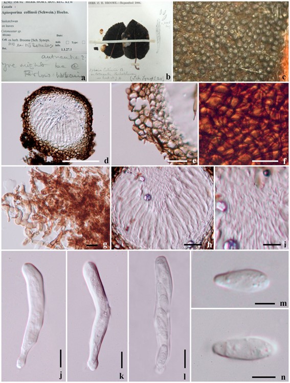
Apiosporina collinsii (Schwein.) Höhn., Sber. Akad. Wiss. Wien, Math.-naturw. Kl., Abt. 1 119: 439 (1910).
≡ Sphaeria collinsii Schwein., Trans. Am. phil. Soc., New Series 4(2): 211 (1832) [1834].
MycoBank number: MB 122234; Index Fungorum number: IF 122234; Facesoffungi number: FoF 06355; Fig. 92
Parasitic on leaves, stems petioles, buds and flowers appearing as black dots like areas. Sexual morph: Ascomata 150–200 high× 150–200 diam. μm ( x̄ = 194 × 195 μm, n= 10) small, gregarious, superficial, globose to subglobose, developing on a dark hyphal mass, easily removed from the substrate, small papillate and ostiolate. Hypha 4 μm wide, pale brown to dark brown, branched, septate. Peridium thinwalled, composed of three layers, dark brown to hyaline cells of textura angularis. Hamathecium comprising 4 μm wide, rare, septate, cellular pseudoparaphyses. Asci 7–9 ×55–65 μm ( x̄ = 7.8 × 62.2 μm, n = 10), 8-spored, bitunicate to narrowly obclavate, cylindrical, with a short, thick, furcate or knob-like pedicel or pedicel lacking. Ascospores 4–6 × 12–15 μm (x̄ = 5.1 ×13.4 μm, n = 10), uni-seriate and partially overlapping, ovoid to narrowly ovoid, hyaline to pale brown, apiosporous, 1-septate near the lower end, barely constricted at the septum, verrucose. Asexual morph: Cladosporium sp. (Sivanesan 1984).
Material examined: Canada, Saskatchwan, on leaves of Cotoneaster sp. (K(M):158702, ex herb. Broome).

Fig. 92 Apiosporina collinsii (K(M):158702). a, b Herbarium specimens. c Ascomata on host substrate. d Cross section of ascoma. e Peridium. f Wall of ascoma composed darkly pigmented cells. g Hyphal mass. h Arrangement of hamathecium. i Pseudoparaphyses. j–l Asci. m, n Ascospores. Scale bars: d = 100 μm, e, g, h = 20 μm, f, i–l = 10 μm, m, n = 5 μm
