Anteaglonium thailandicum Jayasiri & K.D. Hyde, in Jayasiri, Jones, Kang, Promputtha, Bahkali & Hyde, Phytotaxa263(3): 238 (2016)
Index Fungorum Number: IF551984
Holotype: MFLU 16-0471
Etymology: With reference to the country where the specimen was found.
Saprobic on decorticated wood of an unidentified plant. Sexual morph: Hysterothecia 250–430 μm long, 200–250 μm high, 170–200 μm wide, superficial, carbonaceous, black, subglobose to oblong, straight, smooth or slightly striate laterally, with a longitudinal slit, sulcus shallow, gregarious, linear or rarely lying at irregular angles, occurring on a black thin crust, tending to darken the substratum, without KOH extractable pigments. Peridium 34–66 μm thick, carbonaceous, brittle with age, longitudinally striated at the margins, thickened towards apex, base less thick, the inner layer compressed and pallid, the outer layer thickened, comprising pigmented cells of textural angularis. Hamathecium comprising numerous, 1–1.5 μm (n = 30) wide, aseptate pseudoparaphyses, branched above the asci. Asci 45–50 × 3.5–5.5 μm, 8-spored, bitunicate, cylindrical, short pedicellate, obliquely to irregularly uniseriate. Ascospores 6.4–7.8 × 2.4–3.1 μm (x = 7 × 2.8 μm; n = 20), ellipsoid to obovoid, straight, hyaline, smooth-walled, 1-septate, constricted at the septum, guttulate (Fig. 1). Asexual morph: Coelomycetous. Conidiomata 100–150 μm high, 150–170 μm diam., pycnidial, globose, superficial to subperidermal, separate, unilocular, thick-walled, ostiolate. Conidiomata wall 30–50 μm wide, composed of 6–8 layers, with outer 2–3 layers of pale brown and inner 4–5 layers of hyaline cells of texura angularis. Conidiophores long, unbranched, hyaline, formed from the innermost layer of wall cells. Conidiogenous cells 3–5 × 2–3 μm, globose, hyaline, smooth, with a rounded tip. Conidia 3–5 × 2–3 μm (x = 4 × 2.5 μm; n = 20), hyaline, oval to globose, rounded at both ends, aseptate, smooth-walled (Fig. 2).
Culture characters: Colonies on MEA 23 mm diam. after 7 d, raised, with lobate margin; colony two-layered, outer layer white and inwardly black. with asexual structures, finely floccose to woolly aerial mycelium in outer layer. Reverse off-white with middle black.
Material examined: THAILAND. Chiang Rai: Doi Pui, a dead branch of undetermined, 15 June 2014, Subashini C. Jayasiri (MFLU 16-0471, holotype; isotype in KUN); ex-type culture, MFLUCC 14-0816, KUNCC.
Notes:—This species belongs to the genus Anteaglonium because of its morphological features, in particular, the oval to elongate, or globose to subglobose, black, carbonaceous, hysterothecia, which are superficial or sunken in the substrate, cylindric-clavate, short-pedicellate asci, and uniseriate, hyaline, two-celled, small (less than 8 μm long) ascospores. It also differs in the multigene phylogenetic analysis, where it clusters with other Anteaglonium species but is distinct. Anteaglonium globosum resembles A. thailandicum in spore shape and size and intends to darken the substratum, but Anteaglonium globosum differs from A. thailandicum in having globose hysterothecia with a roughened wall, an indistinct slit, short subicula, short tomentum hysterothecial wall and producing a strong green soluble pigment in KOH. Anteaglonium thailandicum lacks a subiculum, has a rough, tomentose and irregular hysterothecial wall and lacks KOH extractable pigments. These character differences and multigene phylogenetic analysis are used to justify the introduction of this new species.
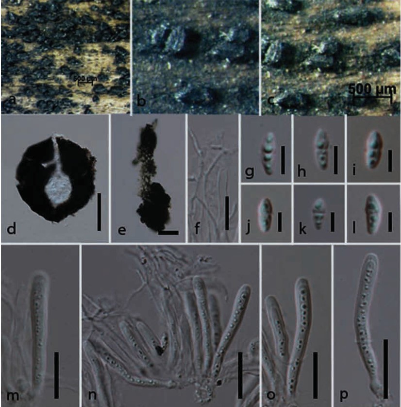
Fig. 1. Anteaglonium thailandicum (holotype). a–c. View of hysterothecia on host surface. d. Section through hysterothecium. e. Peridium. f. Pseudoparaphyses. g–l. Ascospores. m–p. Asci. Scale bars: d=100 μm, e=20 μm, f=10 μm, g–l=5 μm, m–p=20 μm.
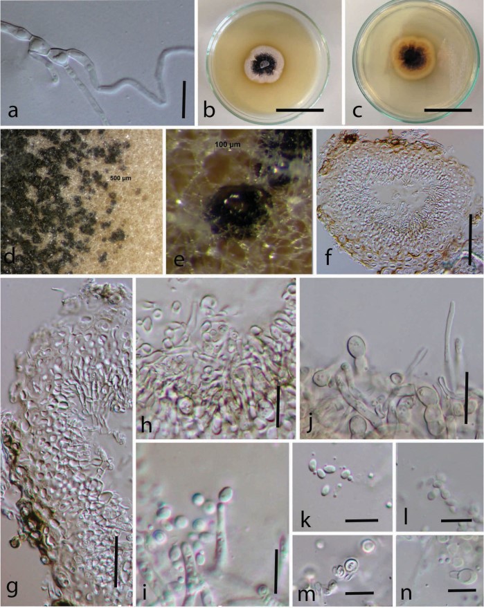
Fig. 2. Asexual morph of Anteaglonium thailandicum from culture. a. Germinated spore. b, c. Culture from above and below. d, e. Fruiting bodies forming in culture. f. Vertical section of conidioma. g. Conidiomata wall. h. Conidiophore. j, i. Conidiogenous cells. k–n. Conidia. Scale bars: a=10 μm, f=50 μm, g–j=20 μm, k–n=5 μm, k=10 μm.