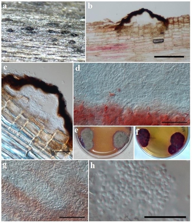
Anteaglonium parvulum (W.R. Gerard) Mugambi & Huhndorf, Syst. Biodiv. 7(4): 462 (2009). Fig. 48
≡ Hysterium parvulum W.R. Gerard, Bull. Torrey bot. Club 5: 40 (1874).
MycoBank number: MB 543261; Index Fungorum number: IF 543261; Facesoffungi number: FoF 01931;
Saprobic on Morinda citrifolia decaying twig. Sexual morph: Undetermined. Asexual morph: Coelomata 100–120 × 170–350 µm, pycnidial, immersed to erumpent, covered by the host periderm, raised at centrre, scattered, brittle, dark brown, homogenously pigmented, interior lined with a slightly red color. Peridium 20–25 µm wide, composed of brown to dark brown cells of textura globulosa, absent at base. Conidiophores and Conidiogenous cell are insignificant or absent. Conidia 2.1–2.7 µm (x̅ = 2.4, n = 20), globose, hyaline, densely present, smooth-walled.
Culture characteristics – Thick Colonies on MEA, 17–20 mm diam. within 7 days at 25°C, short, surface whitish, turning greyish, often with red margin, bottom red.
Material examined – India, Andaman and Nicobar Islands, South Andaman, Kalatan (11˚47’52.6”N 92˚42’50”E), on a twig of Murraya exotica (Rutaceae), 10 August 2016, M. Niranjan & V.V. Sarma PUFNI 17626 (AMH-10075), living culture NFCCI-4375.
GenBank numbers – ITS: MN582759; SSU: MN582763.
Notes – Anteaglonium species, by virtue of having hysterothecoid ascomata and hyaline two- celled ascospores, are similar to Psiloglonium spp., except for the smaller size of their ascomata and ascospores (Boehm et al. 2009a). Anteaglonium abbreviatum, A. globosum, A. latirostrum and A. parvulum were added to the genus by Mugambi & Huhndorf (2009b), A. brasiliense by De Almeidia et al. (2014), A. thailandicum by Jayasiri et al. (2016), A. rubescens by Jaklitsch et al. (2018b) and A. gordoniae by Jayasiri et al. (2019). Asexual morphs were found in two species, A. parvulum and A. thailandicum (Jayasiri et al. 2016) and in A. rubescens (Jaklitsch et al. 2018b) under in vitro conditions. In the present study the asexual morph is reported for the first time from a natural substratum. Cultures in MEA medium show circular growth, apically grey coloured appearance and bottom dark red colour. Red colour on the reverse of agar plates was also reported for A. parvulum by Jayasiri et al. (2016) and A. rubescens in CMD, MEA and PDA media (Jaklitsch et al. 2018b).

Figure 48 – Anteaglonium parvulum (PUFNI 17626, holotype). a Ascomata. b, c Vertical section. d, g Rose pigments in sterile hyphae. e, f Asexual morph in petri plates. h Conidia. Scale bars: b = 200 µm, c, d, g = 50 µm, h = 20 µm.
