Amphisphaeria thailandica Samarak. & K.D. Hyde, in Samarakoon, Liu, Hyde & Promputtha, Phytotaxa 391(3): 212 (2019)
Index Fungorum number: IF 555397; Facesoffungi number: FoF 04977
Etymology – The specific epithet thailandica refers to the country in which the fungus was collected.
Holotype – MFLU 18–0794
Saprobic on a recently dead branch. Sexual morph: Ascomata 210–265 μm high × 410–470 μm diam. (x̅ = 237 × 442 μm, n = 10), immersed, visible as black spots, flat or concave on the host surface, solitary, scattered, subglobose, short, wide papillate, brown, ostiolate. Periphyses 0.9–1.4 μm wide (x̅ = 1.2 μm, n = 20), hyaline, short. Peridium 13.5–30 μm (x̅ = 21 μm, n =15), comprising an inner layer of hyaline cells of textura angularis, the outer layer of reddish brown cells of textura angularis, easily detachable from the host when mature. Paraphyses 5.2–9 μm wide ( x = 6.9 μm, n = 20), hyaline, few, longer than asci, cellular, constricted, septate, guttulate, embedded in a gelatinous matrix. Asci 95–120 × 9.5–16 μm (x̅ = 110 × 12.5 μm, n = 25), 8-spored, unitunicate, cylindrical, with bifurcate pedicel, apically rounded, J-. Ascospores 12.3–15 × 6.9–8.8 μm (x̅ = 13.7 × 7.7 μm, n = 35), uniseriate, subglobose to oval, hyaline bi-guttulate when young, light brown to grayish bi-guttulate at maturity, uniseptate, constricted at the septum, smoothwalled. Asexual morph: Undetermined.
Material examined – Thailand, Phayao, Phu Sang, Doi Phu Nang, on a recently dead branch, 20 July 2017, MC. Samarakoon, SAMC097 (MFLU 18–0794, holotype), ibid., (HKAS 102290, isotype).
Additional sequences – RPB2: MK033640, SSU: MK031922, TUB2: MK033639
Distribution – Thailand
Note – Amphisphaeria thailandica is similar to A. bertiana Fairm., A. paedida (Berk. & Broome) Sacc., A. sorbi Senan. & K.D. Hyde and A. vibratilis (Fuckel) E. Müll in having asci with J-, apical apparatus. Amphisphaeria thailandica can be distinguished from other Amphisphaeria species by having subglobose to oval, hyaline bi-guttulate immature and light brown to grayish bi-guttulate ascospores, lacking a mucilaginous sheath. In addition, A. thailandica possesses a minimum ascospore L/W ratio (1.77) as compared to A. bertiana (2.44), A. paedida (3.1), A. sorbi (2.92) and A. vibratilis (3.3) (Wang et al. 2004, Liu et al. 2015). Amphisphaeria sorbi and A. vibratilis differ from A. thailandica in having a thick mucilaginous sheath around the ascospores. Amphisphaeria depressa Petr. possesses depressed ascomata, which are similar to a few mature ascomata observed in A. thailandica. However, A. depressa differs from A. thailandica in having asci with J+, discoid apical apparatus. A comparison of the 5.8S region of the ITS gene reveals 5.69% bp differences between A. thailandica and A. umbrina (Jeewon & Hyde 2016).
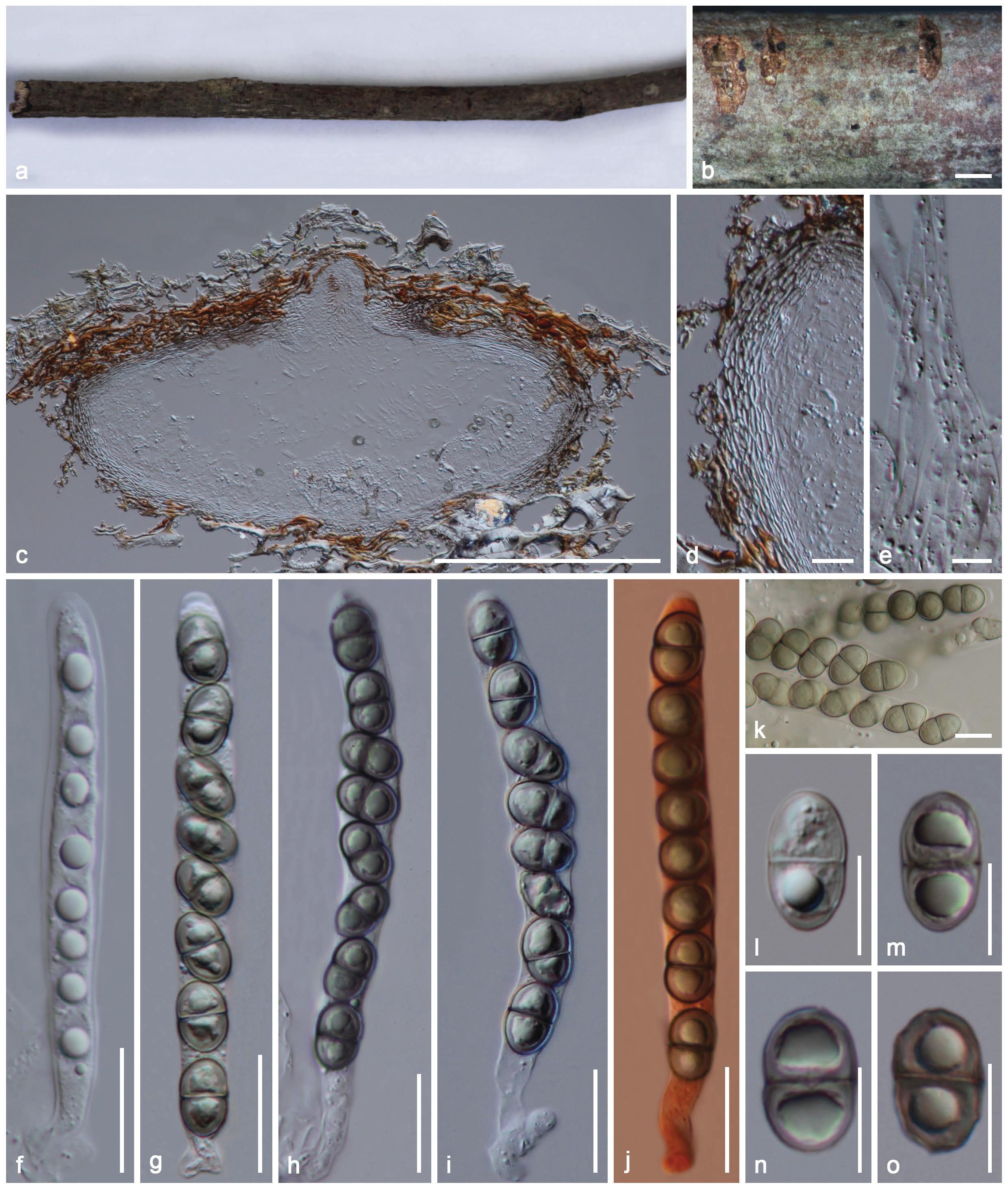
Fig 1. Amphisphaeria thailandica (holotype MFLU 18–0794) a-b. Ascomata on substrate. c. Vertical section of ascoma. d. Peridium. e. Paraphyses. f-j. Asci (j in Congo Red). k-o. Ascospores (k in Melzer’s reagent). Scale bars: b = 1000 μm, c = 200 μm, d, f–j = 20 μm, e, k, o = 10 μm.