Amarenomyces ammophilae (Lasch) O.E. Erikss., Op. bot. 60: 124 (1981)
≡ Sphaeria ammophilae Lasch Flora, Jena 33: 282 (1850)
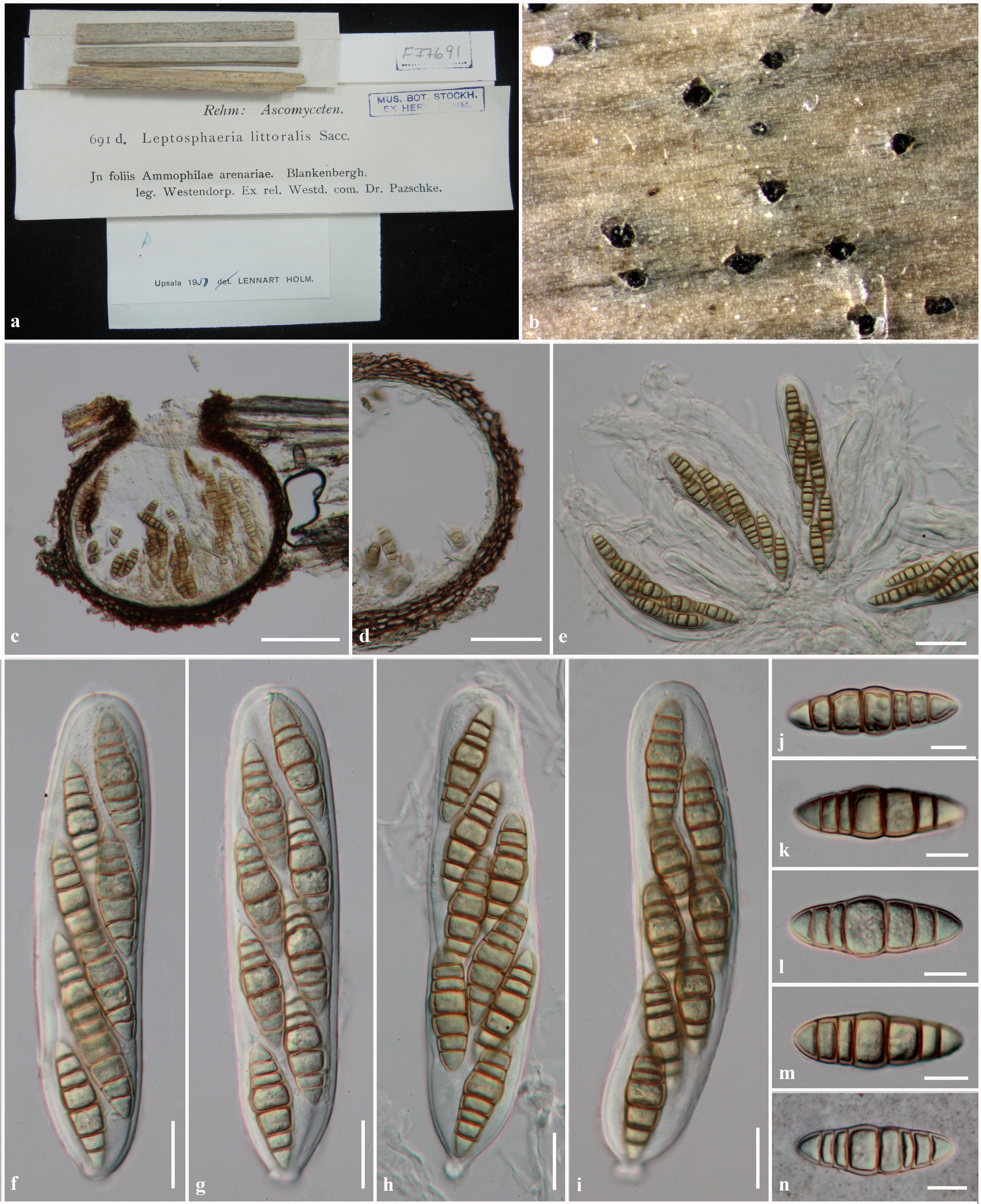
Saprobic on Ammophila. Sexual state: Ascomata 170– 290μm high, 170–285μm diam., scattered, immersed to erumpent through host tissue, visible as small black spots on host surface, uniloculate, globose to subglobose, glabrous, dark brown, ostiole central, with short papilla (50–130μm long). Peridium 7.5–25μm wide, thin-walled, of equal thickness, composed of 3–5 layers of flattened, dark brown, pseudoparenchymatous cells, arranged in textura angularis to textura prismatica. Hamathecium composed of numerous, 1.5–3μm wide, filiform, distinctly septate, frequently anastomosing, narrow cellular pseudoparaphyses, embedded in mucilaginous matrix. Asci (112–)140–160(−173)× (28–)30–35(−37) μm (x = 140×32.4μm, n=25), 8-spored, bitunicate, fissitunicate, broadly cylindrical, subsessile with minute knob-like pedicel, apically rounded, with a well-developed ocular chamber. Ascospores 39–47(−50)× 12–14 μm (x = 43.4 × 13.2 μm, n = 30), overlapping 1–3-seriate, phragmosporous, broadly fusiform, rarely
cylindric-clavate, widest at the middle cell, pale yellowish when immature, becoming yellowish-brown to brown at maturity, 6-septate, slightly constrict at the septum, smooth and thick-walled.
Material examined: BELGIUM, West-Vlaanderen, Blankenbergh, on Ammophila arenaria L. (Poaceae), Westendorp (S-F77691); ITALY, Sardegna, Cagliari, La Maddelena, March 1866, Marcucci (S-F77741).
Notes: Amarenomyces was introduced by Eriksson (1981) to accommodate the single species froma marine grass, which traditionally has been identified as Sphaeria ammophilae Lasch. Eriksson (1981) mentioned that the genus should belong in Botryosphaeriaceae rather than Phaeosphaeriaceae as its morphological characters differed from Phaeosphaeria. Zhang et al. (2009) listed Amarenomyces as a synonym of Phaeosphaeria based on phylogenetic evidence. The generic type of Amarenomyces, A. ammophilae often clusters with other Phaeosphaeria species (Zhang et al. 2009, 2012; Hyde et al. 2013). Thus the earlier name as proposed by Kohlmeyer and Kohlmeyer (1965) and Leuchtmann (1984) was reinstated as Phaeosphaeria ammophilae (Suetrong et al. 2009; Zhang et al. 2009). Several Phaeosphaeria species have previously been transferred to other genera based on the phylogenetic evidence and due to their asexual state. For example Phaeosphaeria nodorum which was transferred to Parastagonospora, as Pa. nodorum. On the other hand, Shoemaker and Babcock (1989b) divided Phaeosphaeria into six subgenera by combining Phaeosphaeria species with similar morphological characters, but differing in ascospore characters, host preference and asexual state (Câmara et al. 2002; Zhang et al. 2012; Quaedvlieg et al. 2013). The asexual state of Phaeosphaeria has been reported as Phaeoseptoria and this is supported by phylogenetic data (Quaedvlieg et al. 2013). Quaedvlieg et al. (2013) transferred Phaeosphaeria species which did not produce Phaeoseptoria asexual states to other genera. Therefore, the genus Amarenomyces is reinstated in this study based on the phylogenetic analysis as A. ammophilae forms a separate clade to the type species, Phaeosphaeria oryzae. Although A. ammophilae has often been reported to be the sexual state of Amarenographium
metableticum (Trail) O.E. Erikss., they are not treated as congeneric here. The presumed link was established as both species occur on the same host in close proximity. There is, however, no sequence data for Amarenographium metableticum and it might be predicted that if both species were host-specific then it would not be surprising that they both occur on the same host. We therefore refrain from combining the taxa until molecular data confirms the link.
Fig. 1 Amarenomyces ammophilae (S-F77691). a Specimens and herbarium label b Ascomata visible on host surface. c Section through ascoma. d Section through peridium. e Asci with cellular pseudoparaphyses. f–i Asci. j–m Ascospores. n Ascospore stained in indian ink. Scale bars: c=100μm, c, d=50μm, e, f, g, h, i=20μm, j, k, l, m, n=10μm