Amarenographium metableticum (Trail) O.E. Erikss., Mycotaxon 15: 199 (1982)
≡ Camarosporium metableticumTrail, Scott. Natural., N.S. 2 (‘8’): 267 (1886)
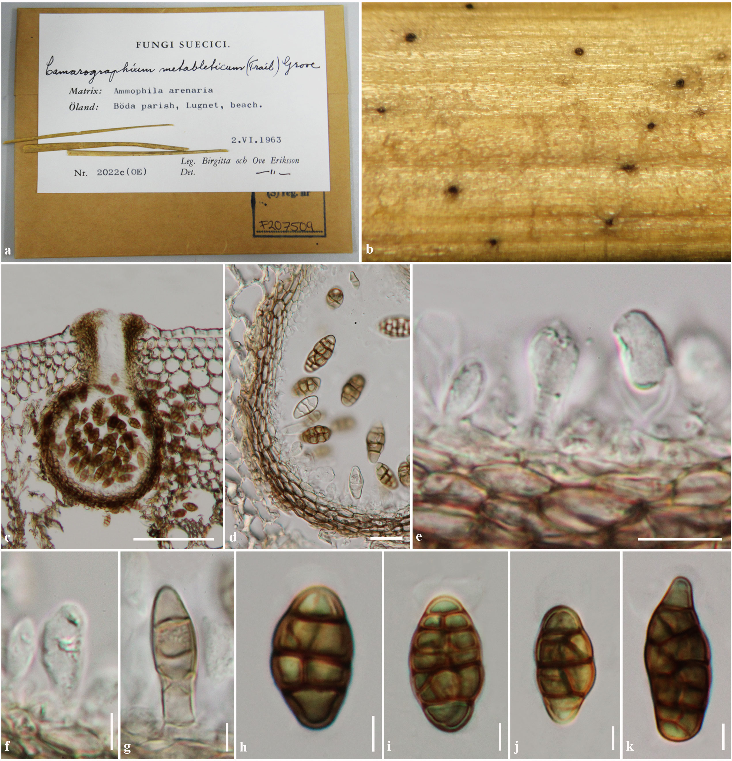
Saprobic on Ammophila. Sexual state: see notes. Asexual state: Conidiomata 160–215.5μm high, 150–270μm diam., pycnidial, scattered, solitary, immersed to erumpent, visible as black spots on host surface, uniloculate, globose to subglobose, glabrous, dark brown, ostiole central, with short to long papilla. Conidiomata walls 13–19μm wide, thinwalled, of equal thickness, composed of 3–5 layers of flattened, brown to dark brown, pseudoparenchymatous cells, arranged in a textura angularis to textura prismatica. Conidiophores arising frombasal cavity of conidioma, mostly reduced to conidiogenous cells, hyaline to brown, septate. Conidiogenous cells (1.5–)3 4(−9)×1.5–2.5(−5) μm (x = 3.9×2.3μm, n=10), holoblastic, phialidic, discrete oblong to ampulliform, hyaline to brown, 1-septate at the base, smooth-walled. Conidia (20–)24–27(−29)×12– 15(−17) μm (x = 25.4×13.4μm, n = 30), solitary, dictyosporous, muriform, ellipsoidal, ovoid, or lageniform, with obtuse ends or sometimes truncate at the basal cell, initially hyaline, becoming brown to yellowish-brown at maturity, mostly 3–4 transverse septa, 1–4 longitudinal septa, with several segments, smooth and thick-walled, with indistinct mucoid appendage.
Material examined: SWEDEN, Öland, Böda parish, Lugnet beach, on Ammophila arenari (L.) Link (Poaceae), 2
June 1963, B. Eriksson & O. Eriksson 2022c (S-F207509)
Fig. 1 Amarenographium metableticum (S-F207509). a Specimens and herbarium label of Amarenographium metableticum b Conidiomata visible on host surface. c Section through conidiomata. d Section through conidiomata wall. e–g Conidiogenous cells. h–k Conidia. Scale bars: c= 100μm, d=20μm, e, h, i, j, k=10μm, f, g=5μm.