Alternaria tillandsiae E.G. Simmons & C.F. Hill, CBS Biodiversity Ser. (Utrecht) 6: 314. (2007). Index Fungorum Number: IF505007
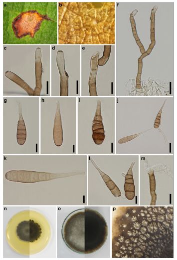
Associated with necrotic leaf lesion of T. grandis. Primary conidiophores up to 175μm× 6–8μm wide in broadest part, simple to branched, straight to slightly curved, septate, slightly constricted at septa, solitary to clustered, hyaline at the tip, apical conidiogenous locus, pale brown to reddish brown at the base, swollen at the base. Conidia (40–)50–65(−90) × 8– 15μm (x = 55 × 11μm, n = 30), ovoid or ellipsoid, with 3–8 transverse septa, and 1–2 longitudinal septa, and 1–2 trans verse segments solitary, pale brown, narrowly ovoid, obclavate, obpyriform, ovoid or ellipsoidal, often with a short conical or cylindrical beak, smooth-walled.
Culture characteristics: Conidia germinating on PDA within 24 h. Colonies on MEA reaching 38–50 mm diam. after 7 days in the dark at 25 °C (x = 28, n= 5), circular, fimbriate, fluffy, convex with papillate surface in the center, flat or effuse at the edge, aerial, dense, grey (4E1) from above, olive brown (4F3) from below.
Habitat: Tillandsia usneoides (Bromeliaceae) (Simmons 2007; Woudenberg et al. 2014) and necrotic leaf lesion on T.grandis leaf (current study).
Known distribution: USA, intercepted in New Zealand (Simmons 2007; Woudenberg et al. 2014) and Thailand (current study).
Material examined: THAILAND, Chiang Rai Province, Muang District, on necrotic leaf lesion of T. grandis, 7 June 2012, M. Doilom, MFLU 15–3421, living culture MFLUCC 12–0383, MKT 056, ICMP 21159, GenBank Accession No: GAPDH: KU764717, ITS: KU144925, RPB2: KU764718, TEF1: KU764719.
Notes: Alternaria tillandsiae strain MFLUCC 12–0383 inthis study was observed on a necrotic leaf lesion in Muang District. No other similar lesions were observed in all collections in this study. Given that the type was collected on Tillandsia usneoides, which is a bromeliad from the USA, and has not been recorded on another host (SMML database accessed 6-Feb-2016) it would seem unlikely that this is the same species. The morphology varies from the type description, however, the type is from a culture, whereas this collection was from host tissues which might explain the morphological differences. The conidia of this collection are shorter and smaller (50–90 × 8–14 μm versus 70–102 × 16–19 μm) than those reported by Simmons (2007) for the type, However, phylogenetic analysis supported this collection as A. tillandsiae. This is the first report of A. tillandsiae from T. grandis in Thailand therefore, details are provided to facilitate identification of this species if it is subsequently recorded again on teak
FIG Alternaria tillandsiae (MFLU 15–3421). a Necrotic lesion host„ surface. b Close-up of colonies on leaf. c–f Conidiophores. g–l Conidia. m Germinated conidiophore. n Colony on MEA after 7 days (above and below views). o Colony on PDA after 3 months (above and below views). p Dendritic (tree-like) crystals produced after 2 weeks on PDA. Scale bars: c–e, i, l, m = 10 μm, f–h, j = 20 μm, k = 5 μm