Agrocybe manihotis Pegler, Kew Bulletin 21 (3): 508 (1968) Fig.11
MycoBank number: MB 325925; Index Fungorum number: IF 325925; Facesoffungi number: FoF 10791;
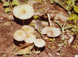
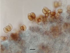
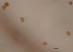
Pileus 3.0 – 6.0 cm in diameter, light buff, shining, slimy, deeply pigmented at the disc, subglobose to convex becoming applanate, , sub- umbonate, smooth, slightly striate at the margin. Flesh moderately thick, pale, changing to brown when bruised . Stipe length 0.7 – 3.0 cm × 0.2– 0.7 cm in diameter, concolorous with the pileus, equal or attenuated towards the apex, hollow, longitudinally striate,pruinose over the entire length, arising from a bulbous base with mycelial strands, veil absent. Lamellae free to adnexed, ferruginous, darkening to umbrinous, crowded, edge white , pruinose. Spores 10.0 – 14.0 × 7.5 – 9.5µm, ovoid to broadly ellipsoid,, rust brown, smooth with a thickened wall, and truncate germ pore, containing one to several guttules.Basidia inflated clavate, 4-spored. Cheilocystidia cylindrical, thin-walled, hyaline present, forming a heteromorphous gill- edge, clavate to cylindric. Pleurocystidia abundant.
Ecology and distribution – Agrocybe manihotis has been reported by Natrajan and Raman 1983 from South India, Kumaresan, et al.2021 have reported this species growing solitary on ground amongst grass from Puducherry, India. Pegler (1977) from East Africa, growing on fallen trunk of trees. Presently, it has been reported to be growing on dung growing gregariously in Mount Abu, Sirohi district of Rajasthan.
Specimens Examined – JNV/Mycl/ 1033/2018, by Reenu Chouhan, 24°53’18.17″N72°50’52.58″E Sirohi, elevation:321 m (1,053 ft), Mount Abu 72.7083°E 24.5925°N Toad rock area.
Note – The morphological characterization of the species described herein are in close resemblance with the description of Agrocybe manihotis described by Pegler 1983 from East Africa except in ————-Although it has been reported from South India by Natrajan and Raman 1983, this is a new report of the taxon from North west India.

a

b

c
Fig.11 a. Fruiting bodies of Agrocybe manihotis .b. Hymenium layer with basidia and spores.c. Spores Scale bar: b = 15 µm, c= 10 µm
