Murispora hawksworthii Wanasinghe, E.B.G. Jones & K.D. Hyde, in Wanasinghe, Jones, Camporesi, Wijayawardene, Mortimer, Xu, Bahkali & Hyde, Cryptog. Mycol. 36(4): 437 (2015)
Facesoffungi Number: FoF01109; Index Fungorum number: IF551561
Etymology: In honour of David Leslie Hawksworth, to celebrate his 70th birthday and his immense contribution to mycology.
Holotype: MFLU 15-2251
Saprobic on dead herbaceous branches of terrestrial habitats. Sexual morph: Ascomata 250-310 μm high 320-380 μm diam. (x̅ = 271.2 × 346.9 μm, n = 10), globose to subglobose, solitary, dark brown to black, superficial, substrate stained purple, fused to the host tissue, ostiolate. ostiole 70-90 μm high 35-50 μm diam. (x̅= 83.3 × 42.4 μm, n = 5), short to papillate, black, smooth, opening to the exterior through the host surface. Peridium 15-25 μm wide at the base, 30-40 μm wide on sides, comprising 3-4 layers of dark brown to black cells of textura angularis, with inner 1-2 layers of cells thin-walled and hyaline. Hamathecium comprising numerous, 1.5-2.5 μm (n=30) wide, filamentous, branched, septate, pseudoparaphyses. Asci 150-200 × 20-28 μm (x̅ = 173.1 × 22.4 μm, n = 30), 8-spored, bitunicate, fissitunicate, cylindric-clavate, shortly pedicellate, thick-walled at the apex, with a minute ocular chamber. Ascospores 25-35 × 8-12 μm (x̅ = 31.6 × 10.4 μm, n = 50), overlapping 1-2-seriate, hyaline when young, becoming pale brown at maturity, ellipsoidal to fusoid, asymmetrical with one side flattened, muriform, with 1-2 longitudinal septa in all cells except end cells, constricted at the septa, conical and narrowly rounded at the ends, guttulate, with a rugged surface, surrounded by a mucilaginous sheath. Asexual morph: Coelomycetous, formed in culture. Conidiomata 1.5-2 mm diam. pycnidial, solitary, dark brown to black, mainly immersed. Pycnidial wall reddish brown cells of textura angularis, with innermost layer thin, hyaline. Conidiophores reduced to conidiogenous cells. Conidiogenous cells phialidic, hyaline, smooth, formed from the innermost layer of pycnidial wall. Conidia (2.5-3.5) × (1.5-2) μm (x̅ = 3.0 × 1.7 μm, n = 50), hyaline, aseptate, guttulate, straight to curved, thin-walled, ellipsoidal.
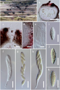
Fig. Murispora hawksworthii (holotype). a. Ascomata on host substrate b. Section of ascoma c. Pseudoparaphyses d. Close up of ostiole e. Peridium f-h. Asci i-k. Ascospores (Note the sheath in k). Scale bars: b = 50 μm, c, e = 10 μm, d, f-h = 20 μm, i-k = 10 μm.
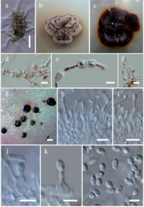
Fig. Murispora hawksworthii Asexual state (holotype). a. Germinating ascospore. b, c. Colonies on PDA (c from below). d. Chlamydospores Intercalary chlamydospores. f. Terminal chlamydospores g. Close up of conidioma. h-k. Immature and mature conidia attached to conidiogenous cell. l. Conidia. Scale bars: a = 20 μm, d-f, h,i = 10 μm, g = 200 μm, j-l = 5 μm.