Cantharellus nigrescens Buyck, Randrianjohany & V. Hofstetter
MycoBank: MB 813240 Facesoffungi number: FoF00991
Etymology: the name refers to the rapidly blackening basidiomata Diagnosis: Can be distinguished from C. congolensis by its more robust stature and frequently fasciculate growth, as well as by its association with Eucalyptus robusta.
Holotype: MADAGASCAR, Talatanivolonondry, in E. robusta plantations along the road to Anzojorobe, 10 February 2006, Buyck & V. Hofstetter 06.197 (PC0084076). Cap up to 15 cm or more, when young very pale, cream to pale yellowish brown or even whitish when remaining covered by dead leaves or other detritus, with appressed, grayishbrownish squamules which become less visible when the cap is expanding, rapidly turning to reddish brown or blackish brown, and finally black. Hymenophore strongly decurrent and well delimited, irregularly and strongly veined-sinuate as well as anastomosing as to almost forming pores, up to 3 mm high, with obtuse edges, pale grayish or isabelline, then turning mouse gray and finally blackish gray to black in an unregular pattern. Stipe short and robust, mostly 20– 30×10–20 mm, subcylindrical or slightly narrowing upwards or even ventricose when young, dirty cream or even pure white when young, quickly staining brown or gray and finally blackening with handling or age, smooth, compact and firm. Flesh very firm and thick beneath the cap center, white to whitish with almost some greenish tinges toward the stipe base, rapidly turning to a dirty brownish gray and finally black when cut. Taste mild to even slightly bitter. Smell weak but typical, of apricot. Spore print with distinct yellowish tinges. Spores ellipsoid, (6)6.5-6.98-7.5(8.1)× (3.7)4-4.35- 4.7(4.8) μm, Q= (1.4)1.5-1.61-1.8(1.9), smooth, filled with one to numerous oily inclusions. Basidia mostly 50-65(74)× 7-8(−9) μm, subcylindrical to weakly clavulate, often sinuate, (3-)5-6-spored; basidiola slender, subcylindrical, sinuate, becoming tardily clavate. Subhymenium filamentous, of slender, strongly septate, hyphal cells, 2-3(4) μm wide like the basidium base. Cystidia none. Pileipellis a loose cutis of ramifying, thin-walled hyphal extremities, measuring mostly 5-8(12) μm diam., terminal cells subcylindrical and hardly differentiated from the subapical ones, but often slightly constricted subterminally and subcapitate, all containing a brown diffuse pigment; no incrusting pigments observed at the surface but present in lower tissues. Clamp connections present everywhere.
Material examined: MADAGASCAR, Central Highlands, Andasibe, along road side with planted eucalypts, 17 February 1997, Buyck, Eyssartier & Moreau 97.547 (PC0084821), near Antananarivo, from E. robusta plantations, 6 February 2006, Buyck & V. Hofstetter 06.166 (PC0084979), 06.176 (PC0084078), Talatanivolonondry, in E. robusta plantations along the road to Anzojorobe, 10 February 2006, Buyck & V. Hofstetter 06.197 (PC0084076 holotype), 06.203 (PC0084980).
Notes: This chanterelle is one of the more common and abundantly fruiting species found in the E. robusta plantations of Madagascar’s Central Highlands. On some occasions, it can literally cover the soil but it is not easily noticed because the pale to dirty or grayish brown cap colour is so similar to the fallen, dry eucalypt leaves lying around it. It is often found in important clusters, frequently more or less aggregated or in very tight groups sitting on mycelial mats. Microscopic examination of specimens collected from different places on the Central Plateau show a remarkable homogeneity among these specimens. During the first years of our fungal inventory of Madagascar, we never saw it for sale, but more recently it is commonly found for sale around and in the capital Antananarivo, perhaps as a result of our collecting trips where people observed us filling baskets of this species. There appears to be no particular reason why local people did not eat this species other than the unappealing aspect and almost instant blackening tissues when cooked. In mainland Africa, the similar C. congolensis is a considered a mediocre edible (Beeli 1928; Buyck 1994; Härkönen et al. 2003). We have prepared and consumed it on several occasions without any further complications. Nevertheless, these blackening species appear to be chemically somewhat different from most other chanterelles as they exhibit remarkable macrochemical reactions: first greenish, then brick red and finally dirty brown with potassium, dark gray with silver nitrate, slowly dark olive green with iron sulfate and green with Guaiac (Eyssartier 2001). Our phylogeny shows it to be very closely related to mainland African collections from miombo woodland that are traditionally identified as C. congolensis Beeli (e.g., Buyck et al. 2000), which is a species originally described from equatorial rain forest. Preliminary sequence data reveal that the blackening chanterelles represent more than just a single species with a large distribution in the whole of tropical Africa, and C. nigrescens is not conspecific with the Central African species of the original description (Buyck & Hofstetter unpubl.). Cantharellus avellaneus Pat. is another Malagasy chanterelle possessing a mouse gray hymenophore, but is definitely a different taxon because of the very elongated spores (see discussion in Buyck et al. 2015). Additional spore measurements for other collections of C. nigrescens give very similar results compared to those made for the holotype: 06.203/ 7–7.65-8.25(9)× (4)4.5-4.75-5 μm Q=1.44- 1.62-2 06.166/ (6.2)6.6-7.15-7.7(8.1)×(3.7)3.8-4.16-4.5(5) μm, Q=(1.5)1.6-1.72-1.8(2) 97.547/ (6)6.5-7.01-7.5(8)×4–4.31-4.75(5) μm Q=1.4- 1.63-1.88 Holotype/ (6)6.5-6.98-7.5(8.1)× (3.7)4-4.35-4.7(4.8) μm, Q=(1.4)1.5-1.61-1.8(1.9)
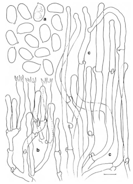
Cantharellus nigrescens (holotype) a Spores b Basidia, basidiola and subhymenial cells c Hyphal extremities of the pileipellis. Scalebars=10μm,butonly5μm for spores. Drawings B. Buyck