Anthostomella thailandica Daranagama & K.D. Hyde, in Hyde et al., Fungal Diversity 80: 213 (2016)
Index Fungorum number: IF 552287; Facesoffungi number: FoF 02419
Etymology – ‘‘thailandica’’ refers to the country it was first collected.
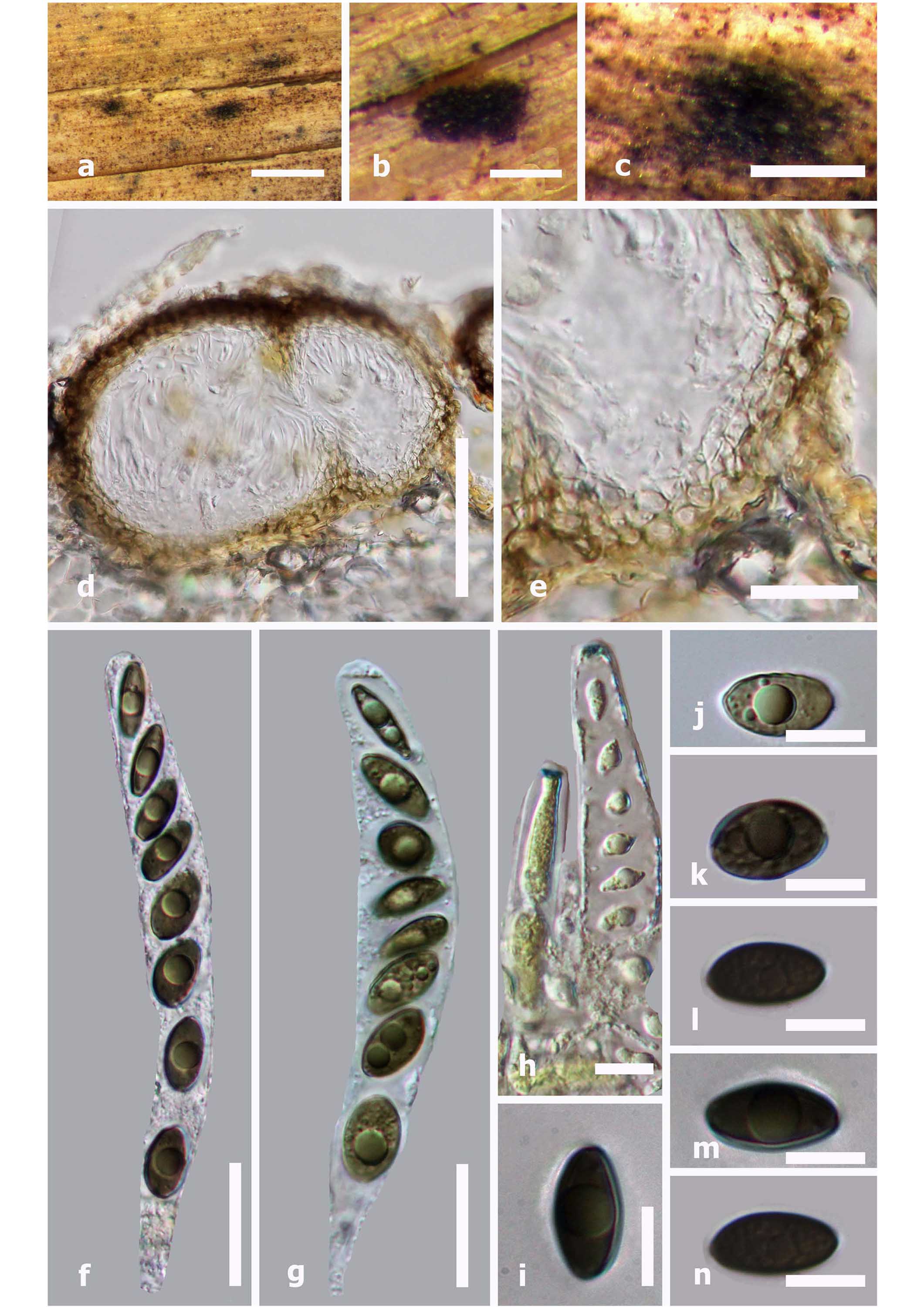
Saprobic on leaves of grasses. Sexual morph: Ascomata 150–200 × 140–155 μm (x̄ = 167 × 148 μm, n = 10), immersed, visible as black, irregular areas, coriaceous, solitary, sometimes clustered in groups of two, scattered, in cross-section subglobose. Ostiole inconspicuous, without conspicuous papilla. Peridium 14–22 μm wide (x̄ = 17 μm, n = 10), comprising two cell layers, outwardly comprising thin-walled, loosely arranged, light brown cells of textura globosa and inwardly comprising a few layers of thin-walled, hyaline cells of textura angularis. Paraphyses 4.2–6.5 μm wide at the base (x̄ = 4.9 μm, n = 30), as long as asci, few, filamentous, septate. Asci 87–118 × 14.1–17.8 μm (x̄ = 92.6 × 15.2 μm, n = 20), 8-spored, unitunicate, cylindrical-clavate, apedicellate, with conspicuous, J+, discoid apical ring, 3.1–3.8 × 1.1–1.9 μm (x̄ = 3.5 × 1.5 μm, n = 20). Ascospores 12–17 × 5.5–9 μm (x̄ = 15.4 9 7.2 μm, n = 20), uni-seriate, equilateral ellipsoidal, with broad ends, olivaceous, smooth-walled, with one large, central guttule, without germ slit. Asexual morph: Undetermined.
Culture characteristics – Colonies on OA 9 cm diam. after 4 weeks at 27 ºC, white at the margins, pale yellowish at the center, reverse yellowish to cream, colony azonate, cottony appearance, dense.
Material examined – THAILAND, Chiang Rai Province, Mae Fah Luang University garden, on dead leaves of grass, 12 August 2015, D.A. Daranagama, KM24 (MFLU 16-0971, holotype), ibid., HKAS, isotype; ex-type living culture, MFLUCC 15-0017.
Notes – Our collection is reminiscent of both A. zongluensis K.D. Hyde and A. consanguinea (Ces.) Sacc. However, A. zongluensis has ascospores with more parallel sides, minutely verruculose walls and ascomata with periphysate ostiolar canals (Lu and Hyde 2000). Ascospores of A. thailandica are similar in size to those of A. consanguinea (Ces.) Sacc. However, ascospores of A. consanguinea have verruculose walls and a short, straight germ slit, while those in A. thailandica have a smooth wall and lack germ slits. In the phylogenetic analysis A. thailandica clusters in a separate clade with A. formosa Kirschst., A. conorum (Fuckel) Sacc., A. rubicola Speg. ex Sacc. & Trotter and A. obesa Daranagama, E. Camporesi & K.D. Hyde.
Fig 1. Anthostomella thailandica (holotype). a–c Appearance of ascomata on the host surface. d Ascoma in cross-section. e Peridium. f, g Mature asci. h Apical ring bluing in Melzer’s reagent. i–n Immature-mature ascospores. Scale bars a = 1 mm , b–c = 500 μm , d = 100 μm , e–g = 20 μm , h = 5 μm , i–n = 10 μm.